
Foundations
BDNF explained: TrkB signalling, how it is measured, and what a rodent BDNF result means
CL
· 10 min read
A summary of published research, with citations. It states no amount, schedule or route, and makes no claim about what this compound does in a person. Supplied for laboratory research use only.
On this page · 7 sections
Brain-derived neurotrophic factor (BDNF) is a growth-factor protein of the neurotrophin family. It supports neuron survival and synaptic plasticity, mainly by activating the TrkB receptor. In research it is measured in several unrelated ways, as messenger RNA, as protein in brain tissue, as receptor activation, or in blood. These are not interchangeable. Serum BDNF in particular is dominated by platelets, and mice have almost none in serum. When a rodent paper on Semax or Selank reports a BDNF change, it is reporting a molecular endpoint, not a cognitive outcome.
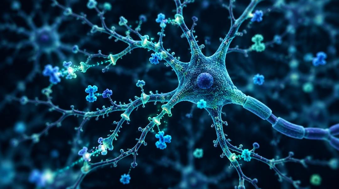
What is BDNF?
The first neurotrophic factor, nerve growth factor (NGF), was known for decades before a second was found. In 1982 Barde, Edgar and Thoenen purified a protein from pig brain that kept cultured embryonic sensory neurons alive and growing. Its molecular weight was about 12,300, it was antigenically and functionally distinct from NGF, and about one microgram was obtained from 1.5 kg of brain (PMID 7188352). The gene was cloned in 1989 (PMID 2779653). BDNF, NGF, neurotrophin-3 and neurotrophin-4 now make up the mammalian neurotrophin family.
That purification yield is worth noticing. BDNF is a scarce, potent signalling protein, and it is made locally and released at synapses. Its biology lies in where and when it is released, which a single bulk concentration captures poorly.
How does BDNF signal through TrkB?
BDNF's main receptor is TrkB, a receptor tyrosine kinase. That makes it a different class from the G protein-coupled receptors behind most peptide hormones on this site. Binding brings two receptor molecules together, and each adds phosphate groups to tyrosine residues on the other, which starts the signal inside the cell.
Klein and colleagues showed in 1991 that BDNF, and neurotrophin-3 but not NGF, rapidly phosphorylated the TrkB receptor protein. In their assays BDNF produced responses at least two orders of magnitude stronger than neurotrophin-3 (PMID 1649702). TrkB is mostly confined to the nervous system, which fits the place where BDNF does most of its work.
There is a second receptor, and it can push the other way. BDNF is made as a longer precursor, proBDNF, which is cut to release the mature protein. Lee and colleagues showed that proneurotrophins are not inert precursors. They preferentially activate the p75 neurotrophin receptor and can drive cell death, whereas the mature forms activate Trk receptors and support survival (PMID 11729324). An assay that does not tell proBDNF from mature BDNF is adding together two molecules that can have opposite effects.
Release matters as much as amount. Egan and colleagues studied a common human variant, val66met, in the pro-region of BDNF. Neurons carrying the met form released less BDNF in response to activity, while their constant background release was unchanged. In human volunteers the met allele was associated with poorer episodic memory and differences in hippocampal function (PMID 12553913). That result is about activity-dependent release at the right place, which no blood test measures.
How is BDNF measured?
| What is measured | Typical method | What it tells you | What it does not |
|---|---|---|---|
| BDNF mRNA, sometimes by exon | Quantitative PCR on tissue | The gene is being transcribed more or less in that tissue | How much protein is made, released or active |
| BDNF protein in brain tissue | ELISA or western blot on homogenate | Total BDNF in that region at that moment | Where it sits, whether it is pro- or mature, whether it is released |
| TrkB phosphorylation | Phospho-specific antibodies | Receptor activation in that tissue | Which downstream effects follow |
| Serum BDNF | ELISA after clotting | Mostly BDNF released from platelets during clotting | Brain BDNF directly |
| Plasma BDNF | ELISA on anticoagulated blood | Free circulating BDNF, much lower and sensitive to handling | Brain BDNF directly |
Each row is a different quantity. A paper that measures one and writes "BDNF" without saying which has left out the most important fact in its methods.
Why is serum BDNF such a difficult proxy for the brain?
Most blood BDNF lives in platelets. Fujimura and colleagues found substantial BDNF stored in human platelets and released when platelets were activated by thrombin, collagen or shear stress. Platelet-poor plasma contained little (PMID 12008958). Platelets had no detectable BDNF mRNA and their precursor cell lines had very little BDNF, so platelets appear to collect it from the circulation rather than make it. Serum is made by letting blood clot, which activates platelets, so serum BDNF largely reflects what platelets were holding and how completely they released it.
Plasma is much lower and tracks platelet release. Radka and colleagues found high BDNF in human and rat serum, up to 50-fold less in citrated plasma, and a strong correlation (r = +0.86) between plasma BDNF and serotonin, another substance released by activated platelets (PMID 8869564). Their reading was that plasma BDNF comes from platelets breaking open, and that truly free circulating levels are negligible.
Mice, the main model, have no serum BDNF to measure. In the same study mouse serum contained no detectable BDNF, even though mouse brain contained plenty. Klein and colleagues confirmed it was undetectable in mouse blood (PMID 20604989). A blood BDNF result therefore cannot be checked against brain levels in the species most mechanism work uses.
Where blood and brain do correlate, the link is partial. Klein and colleagues found positive correlations between whole-blood and hippocampal BDNF in rats (r² = 0.44) and between plasma and hippocampal BDNF in pigs (r² = 0.41), and concluded that blood levels reflect brain levels (PMID 20604989). That is a real finding, and it is worth stating fairly. An r² near 0.4 also means more than half the variation in blood BDNF is unrelated to the hippocampal measure. BDNF can cross the blood-brain barrier through a saturable transport system in mice (PMID 9886678), so blood and brain are not sealed off from each other, but crossing in both directions makes the source of a blood signal harder to identify.
The assays disagree with each other. Polacchini and colleagues ran sera from 40 healthy adults through six commercial kits. Inter-assay variation ranged from 5% to 20%, mostly higher than the manufacturers claimed. Two kits recognised only mature BDNF while the others detected both pro- and mature forms (PMID 26656852). Two studies using different kits may not be measuring the same molecule.
In short, serum BDNF can carry some information about the brain in some species. It is heavily shaped by platelet biology and sample handling, it cannot be validated in mice, and it differs by assay. It is a weak stand-in for BDNF where it acts.
What does "raises BDNF" mean in a rodent paper?
Semax and Selank are the catalogue peptides whose literature most often reports BDNF.
For Semax, Dolotov and colleagues reported that a single exposure in rats produced a maximal 1.4-fold rise in hippocampal BDNF protein, a 1.6-fold rise in TrkB phosphorylation, and threefold and twofold rises in exon III BDNF mRNA and TrkB mRNA. The treated animals also showed more conditioned avoidance responses (PMID 16996037). That is a well-specified study. It measured three different quantities in one brain region, at chosen time points, in rats. What it shows is that the BDNF/TrkB system in rat hippocampus responded. It does not show a cognitive effect in people.
For Selank, a rat study points the other way. Kolik and colleagues gave rats ethanol as their only fluid for 30 weeks. Ethanol raised BDNF content in the hippocampus and frontal cortex, and Selank prevented that increase, which the authors presented alongside better object-recognition performance (PMID 31625062). There the favourable reading attached to BDNF not rising.
Set side by side, the two studies show that a BDNF change has no fixed meaning. It means whatever the model and its other endpoints make of it. "Raises BDNF" is a description of a molecular measurement in a tissue, and the direction that counts as good depends on the experiment. Neither Semax nor Selank is approved by Health Canada, the FDA or the EMA, and their clinical literature comes largely from Russia. The two literatures are compared in Semax vs Selank, with more depth in Semax and stroke research and Selank and the stress response.
A checklist for BDNF claims
- Which BDNF? mRNA, tissue protein, receptor activation, serum or plasma. Pro, mature, or both.
- Which tissue and species? A hippocampal change in a rat is not a serum change in a person.
- Which assay? Kits differ in what form they detect and how reproducible they are.
- Is there a functional endpoint, and was it blinded? A molecular change without one is a mechanism, not an outcome. See how to read a peptide study.
Nothing here is medical, dosing, or therapeutic guidance. For other explainers in this series, see cardiolipin explained and the HPG axis and kisspeptin.
Supplied here for laboratory use: Semax and Selank. The laboratory's certificate for each lot is linked from its product page. For laboratory research use only.
Frequently asked questions
What receptor does BDNF act on? Mainly TrkB, a receptor tyrosine kinase found mostly in the nervous system. In the 1991 study that identified it, BDNF activated TrkB far more strongly than neurotrophin-3. The precursor form, proBDNF, preferentially activates a different receptor, p75, which can promote cell death, so the balance between the pro and mature forms matters.
Does serum BDNF reflect brain BDNF? Only partly. Most serum BDNF is released from platelets during clotting, plasma levels are far lower, and mice have no detectable serum BDNF. In rats and pigs, blood and hippocampal BDNF correlated with r² values around 0.4, which leaves most of the variation unexplained. Assays also differ in which forms of BDNF they detect.
Is BDNF mRNA the same as BDNF protein? No. mRNA shows the gene being transcribed, and protein is what is actually made and can be released. In the Semax rat study, exon III mRNA rose threefold while tissue protein rose 1.4-fold. Both are informative, neither measures release at the synapse, and a paper should say which one it measured.
If a peptide raises BDNF in rats, does it affect memory in people? That does not follow. A BDNF change in rat hippocampus is a molecular endpoint in one species and one tissue. Whether it carries over to people, and whether it changes anything functional, needs separate, controlled human evidence. One Selank study even framed preventing a BDNF rise as the favourable result.
Selected Research References
- Barde YA, Edgar D, Thoenen H. Purification of a new neurotrophic factor from mammalian brain. EMBO Journal. 1982;1:549-553. pubmed.ncbi.nlm.nih.gov/7188352
- Klein R, Nanduri V, Jing SA, et al. The trkB tyrosine protein kinase is a receptor for brain-derived neurotrophic factor and neurotrophin-3. Cell. 1991;66:395-403. pubmed.ncbi.nlm.nih.gov/1649702
- Lee R, Kermani P, Teng KK, Hempstead BL. Regulation of cell survival by secreted proneurotrophins. Science. 2001;294:1945-1948. pubmed.ncbi.nlm.nih.gov/11729324
- Egan MF, Kojima M, Callicott JH, et al. The BDNF val66met polymorphism affects activity-dependent secretion of BDNF and human memory and hippocampal function. Cell. 2003;112:257-269. pubmed.ncbi.nlm.nih.gov/12553913
- Fujimura H, Altar CA, Chen R, et al. Brain-derived neurotrophic factor is stored in human platelets and released by agonist stimulation. Thrombosis and Haemostasis. 2002;87:728-734. pubmed.ncbi.nlm.nih.gov/12008958
- Radka SF, Holst PA, Fritsche M, Altar CA. Presence of brain-derived neurotrophic factor in brain and human and rat but not mouse serum detected by a sensitive and specific immunoassay. Brain Research. 1996;709:122-301. pubmed.ncbi.nlm.nih.gov/8869564
- Klein AB, Williamson R, Santini MA, et al. Blood BDNF concentrations reflect brain-tissue BDNF levels across species. International Journal of Neuropsychopharmacology. 2011;14:347-353. pubmed.ncbi.nlm.nih.gov/20604989
- Polacchini A, Metelli G, Francavilla R, et al. A method for reproducible measurements of serum BDNF: comparison of the performance of six commercial assays. Scientific Reports. 2015;5:17989. pubmed.ncbi.nlm.nih.gov/26656852
- Dolotov OV, Karpenko EA, Inozemtseva LS, et al. Semax, an analog of ACTH(4-10) with cognitive effects, regulates BDNF and trkB expression in the rat hippocampus. Brain Research. 2006;1117:54-60. pubmed.ncbi.nlm.nih.gov/16996037
- Kolik LG, Nadorova AV, Antipova TA, et al. Selank, Peptide Analogue of Tuftsin, Protects Against Ethanol-Induced Memory Impairment by Regulating of BDNF Content in the Hippocampus and Prefrontal Cortex in Rats. Bulletin of Experimental Biology and Medicine. 2019;167:641-644. pubmed.ncbi.nlm.nih.gov/31625062
All products sold by NovaEvum are for research and laboratory use only. They are not intended for human or animal consumption, diagnosis, treatment, cure, or prevention of any disease.