
Testing, quality and handling
Endotoxin Testing for Peptides: LAL, Recombinant Factor C and What Purity Misses
NB
· 9 min read
A summary of published research, with citations. It states no amount, schedule or route, and makes no claim about what this compound does in a person. Supplied for laboratory research use only.
On this page · 8 sections
- What is endotoxin, and how does it get into a synthetic peptide?
- Why doesn't a purity figure capture endotoxin?
- How does the LAL test work?
- What is recombinant factor C, and is it equivalent?
- What does an EU figure mean?
- What is interference, and why can a peptide hide endotoxin?
- What should an endotoxin result on a certificate include?
- Frequently asked questions
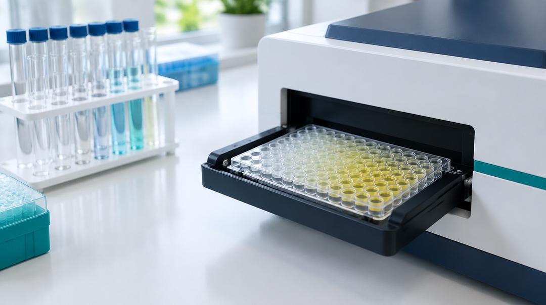
Endotoxin testing for peptides measures bacterial lipopolysaccharide, a potent immune stimulant that a synthetic peptide can pick up from water, glassware and handling even though no bacteria were used to make it. The standard methods are Limulus amebocyte lysate (LAL) assays, in gel-clot, turbidimetric or chromogenic form, and the animal-free recombinant factor C assay. Results are reported in endotoxin units (EU), usually per milligram. HPLC purity cannot detect endotoxin at all, so a peptide can show an excellent chromatogram and still carry enough to dominate a cell or animal experiment.
What is endotoxin, and how does it get into a synthetic peptide?
Endotoxin is lipopolysaccharide (LPS), a component of the outer membrane of Gram-negative bacteria. Raetz and Whitfield describe its three parts: a hydrophobic lipid A domain, which is the endotoxic principle, a core oligosaccharide, and a variable outer polysaccharide, the O-antigen. Animal cells sense lipid A through the receptor TLR4 (PMID 12045108). That receptor is the reason endotoxin matters to researchers: trace amounts can switch on inflammatory signalling in many cell types.
A synthetic peptide is made by chemistry, not fermentation, so it does not start with endotoxin. It acquires it from what it touches. Water systems are the usual source, because Gram-negative bacteria grow readily in them and shed LPS whether alive or dead. Buffers, chromatography columns, glassware, plastics and hands add more. LPS is heat-stable and is not removed by filtration through ordinary sterilising filters, so a solution can be sterile and still contaminated. Sterility and endotoxin are separate questions, a point also made in BPC-157 stability after reconstitution. Synthesis-related impurities, by contrast, are predictable from the chemistry, as set out in solid-phase peptide synthesis explained.
Why doesn't a purity figure capture endotoxin?
HPLC purity is the main peak's share of material absorbing ultraviolet light at the detection wavelength. Lipopolysaccharide has no useful chromophore there, is present at trace mass, and would not elute as a sharp peak if it did. Mass spectrometry is not configured to look for it either. An immaculate chromatogram is therefore silent on endotoxin, as HPLC vs LC-MS peptide testing explains for detector-invisible material generally.
The consequence is not hypothetical. Weinstein and colleagues used an LAL assay to screen commercial- and pharmaceutical-grade reagents used in microglia research and found significant LPS contamination in several commercial-grade proteins, phospholipids and synthetic peptide preparations, but not in pharmaceutical-grade recombinant proteins. Polymyxin B, which neutralises LPS, inhibited the microglia-activating potential of a number of the commercial-grade proteins (PMID 17910052). Naot and colleagues traced an interferon-like gene signature produced by a protein marketed as LPS-free to likely LPS contamination (PMID 22374965). In both cases the contaminant produced an effect that looked like the test substance's biology.
How does the LAL test work?
The assay borrows the clotting system of the Atlantic horseshoe crab, Limulus polyphemus. Levin and Bang showed in the 1960s that the crab's blood cells, amebocytes, contain a clottable protein that coagulates in response to endotoxin (PMID 5690028). A lysate of those cells is the reagent.
The mechanism is an enzyme cascade. Endotoxin activates factor C, which activates factor B, which converts a proclotting enzyme into clotting enzyme, which cleaves the protein coagulogen into coagulin, which gels. Each step amplifies the last, which is why the assay is so sensitive. A second entry point, factor G, is triggered by (1→3)-β-D-glucans from fungi and cellulose materials rather than by endotoxin; Bolden and colleagues note that lysate reagents detect this alternate pathway (PMID 32817324), which is a known source of false positives unless the reagent blocks it.
Three read-outs are in use:
| Format | What is measured | Result type | Notes |
|---|---|---|---|
| Gel clot | Whether a firm gel forms after incubation, at a series of dilutions | Pass/fail at the reagent's labelled sensitivity, or semi-quantitative by endpoint dilution | Simple, the historical reference method, coarse |
| Turbidimetric (kinetic or end-point) | Rise in turbidity as coagulin forms | Quantitative, against a standard curve | Sensitive to sample colour or haze |
| Chromogenic (kinetic or end-point) | Colour released when clotting enzyme cleaves a synthetic substrate | Quantitative, against a standard curve | Sensitive to coloured samples and to enzyme inhibitors |
| Recombinant factor C | Fluorescence when recombinant factor C, activated by endotoxin, cleaves a substrate | Quantitative, against a standard curve | No factor G pathway; no horseshoe crab blood |
What is recombinant factor C, and is it equivalent?
Recombinant factor C (rFC) reproduces only the first enzyme of the cascade, made by recombinant expression, with a fluorogenic substrate in place of the clotting chain. Because the factor G branch is absent, it responds to endotoxin more selectively. Bolden and colleagues reviewed comparisons and found a high degree of correlation between rFC and lysate tests, concluding that the recombinant methods are comparable, while also noting that rFC removes the need to bleed horseshoe crabs (PMID 32817324).
Comparable is not identical. Abate and colleagues tested LPS from a range of bacterial species and found that the rFC assay detected most structures in picogram quantities, with no difference from LAL assays for LPS from E. coli, Burkholderia cepacia and Salmonella, but significantly different reactivity for LPS from Klebsiella pneumoniae, Serratia marcescens, Bordetella pertussis and Pseudomonas aeruginosa. Only the rFC assay correlated significantly with IL-6 release from a human monocytic cell line (PMID 28693666). The species source of a contamination can therefore affect what each assay reports.
What does an EU figure mean?
An endotoxin unit is a unit of biological activity, not mass. It is defined by comparison with a reference standard endotoxin, and every quantitative assay is calibrated against a control standard traceable to that reference. Because LPS from different bacteria differs in potency, an EU value describes how much activity the sample shows in that assay relative to the standard, not how many nanograms of LPS are present.
For a peptide, results are usually reported per milligram of material. Two points help in reading them. A "less than" result, such as below a stated EU/mg, is bounded by the assay's sensitivity and the dilution needed to overcome interference, so the limit itself is informative: a high limit means the test could not look closely. And the figure is per milligram of the material tested, which, as peptide net content vs purity explains, is not the same as per milligram of peptide.
Pharmacopoeias set endotoxin limits for licensed medicines by product. Research material has no such limit. The relevant threshold for laboratory work is what the experimental system tolerates, which for sensitive immune cells can be very low and is worth establishing with an LPS standard in the same system.
What is interference, and why can a peptide hide endotoxin?
An assay can be inhibited or enhanced by the sample matrix. Extremes of pH, chelators, high salt, some proteases and some proteins all interfere. The standard safeguard is a positive product control: the sample is spiked with a known amount of endotoxin and the recovery is measured. If the spike is not recovered within the method's acceptance range, the result for the unspiked sample cannot be trusted, and the sample is diluted and retested, up to the maximum dilution the assay's sensitivity allows.
Peptides have a specific way to interfere. Many cationic, amphipathic peptides bind lipid A; polymyxin B, the LPS neutraliser in the Weinstein study, is itself such a peptide (PMID 17910052). A peptide that binds LPS can mask it from the assay while not necessarily neutralising it in a biological system.
A related phenomenon in biologic formulations, low endotoxin recovery, involves endotoxin spiked into undiluted product becoming progressively less detectable during a hold period. Reich and colleagues found that masking depended on the endotoxin source, so that susceptibility could not be predicted (PMID 30781342), and Cao and colleagues reviewed the contributions of protein ingredients and excipients such as chelators and surfactants (PMID 34407207). Most peptide research material has no formulation of that kind, but the lesson transfers: a clean result is only as good as its spike recovery.
What should an endotoxin result on a certificate include?
The method (gel clot, kinetic turbidimetric, kinetic chromogenic or rFC), the result in EU/mg with its reporting limit, the dilution tested, and evidence of a valid positive product control. Endotoxin testing is a separate commission from purity and identity, which is why third-party peptide testing in Canada lists it on its own row, and why what a certificate of analysis establishes calls it the test most worth asking about for biological work. A certificate that omits it has said nothing about endotoxin, positive or negative. Nothing here is medical, dosing, or therapeutic guidance.
Frequently asked questions
Can a synthetic peptide contain endotoxin if no bacteria were involved in making it? Yes. Endotoxin comes from Gram-negative bacteria in water systems, buffers, equipment and handling, not only from fermentation. It survives heat and passes ordinary sterilising filters, so a chemically synthesised, filtered peptide can still carry it. Weinstein and colleagues found LPS in commercial-grade synthetic peptide preparations screened by LAL assay.
Is a sterile peptide also endotoxin-free? No. Sterility means no viable microorganisms; endotoxin is a molecule shed by bacteria, alive or dead. Filtration removes cells but not LPS, and ordinary heat sterilisation does not destroy it. The two are tested separately, by different methods, and one result says nothing about the other.
Is the recombinant factor C test as good as LAL? Reviews comparing them find a high degree of correlation, and rFC avoids false positives from the glucan-sensitive factor G pathway. Agreement is not perfect for every bacterial source: Abate and colleagues found different reactivity between rFC and LAL for LPS from several species. Either method needs a valid spike-recovery control.
Why would a peptide interfere with the endotoxin assay? Some peptides, especially cationic and amphipathic ones, bind lipid A, which can hide endotoxin from the assay. Others alter pH or inhibit the cascade enzymes. A positive product control, where known endotoxin is added to the sample and must be recovered, is how a laboratory shows the result is valid.
Selected Research References
- Raetz CR, Whitfield C. Lipopolysaccharide Endotoxins. Annual Review of Biochemistry. 2002;71:635-700. pubmed.ncbi.nlm.nih.gov/12045108
- Levin J, Bang FB. Clottable Protein in Limulus; Its Localization and Kinetics of Its Coagulation by Endotoxin. Thrombosis et Diathesis Haemorrhagica. 1968;19(1):186-197. pubmed.ncbi.nlm.nih.gov/5690028
- Weinstein JR, Swarts S, Bishop C, et al. Lipopolysaccharide Is a Frequent and Significant Contaminant in Microglia-Activating Factors. Glia. 2008;56(1):16-26. pubmed.ncbi.nlm.nih.gov/17910052
- Naot D, Williams GA, Lin JM, et al. Evidence That Contamination by Lipopolysaccharide Confounds In Vitro Studies of Adiponectin Activity in Bone. Endocrinology. 2012;153(5):2076-2081. pubmed.ncbi.nlm.nih.gov/22374965
- Bolden J, Knutsen C, Levin J, et al. Currently Available Recombinant Alternatives to Horseshoe Crab Blood Lysates: Are They Comparable for the Detection of Environmental Bacterial Endotoxins? A Review. PDA Journal of Pharmaceutical Science and Technology. 2020;74(5):602-611. pubmed.ncbi.nlm.nih.gov/32817324
- Abate W, Sattar AA, Liu J, et al. Evaluation of Recombinant Factor C Assay for the Detection of Divergent Lipopolysaccharide Structural Species and Comparison with Limulus Amebocyte Lysate-Based Assays and a Human Monocyte Activity Assay. Journal of Medical Microbiology. 2017;66(7):888-897. pubmed.ncbi.nlm.nih.gov/28693666
- Reich J, Weyer FA, Tamura H, et al. Low Endotoxin Recovery: Masking of Naturally Occurring Endotoxin. International Journal of Molecular Sciences. 2019;20(4):838. pubmed.ncbi.nlm.nih.gov/30781342
- Cao Y, Zhang Y, Qiu F. Low Endotoxin Recovery and Its Impact on Endotoxin Detection. Biopolymers. 2021;112(11):e23470. pubmed.ncbi.nlm.nih.gov/34407207
All products sold by NovaEvum are for research and laboratory use only. They are not intended for human or animal consumption, diagnosis, treatment, cure, or prevention of any disease.