
Testing, quality and handling
Does Shaking Damage Peptides? What Agitation Does and How It Is Detected
NB
· 10 min read
A summary of published research, with citations. It states no amount, schedule or route, and makes no claim about what this compound does in a person. Supplied for laboratory research use only.
On this page · 7 sections
Shaking can damage some peptides, but not in the way usually imagined. The main stress is not shear force; it is the air–liquid interface that agitation keeps creating. Peptide molecules adsorb there, rearrange and can nucleate aggregates, while foam and bubbles multiply the exposed surface. How much harm results depends on the sequence, concentration, formulation, fill volume and container. Amphipathic, self-associating and fibril-forming peptides are most at risk, and the damage is often invisible to a routine purity chromatogram.
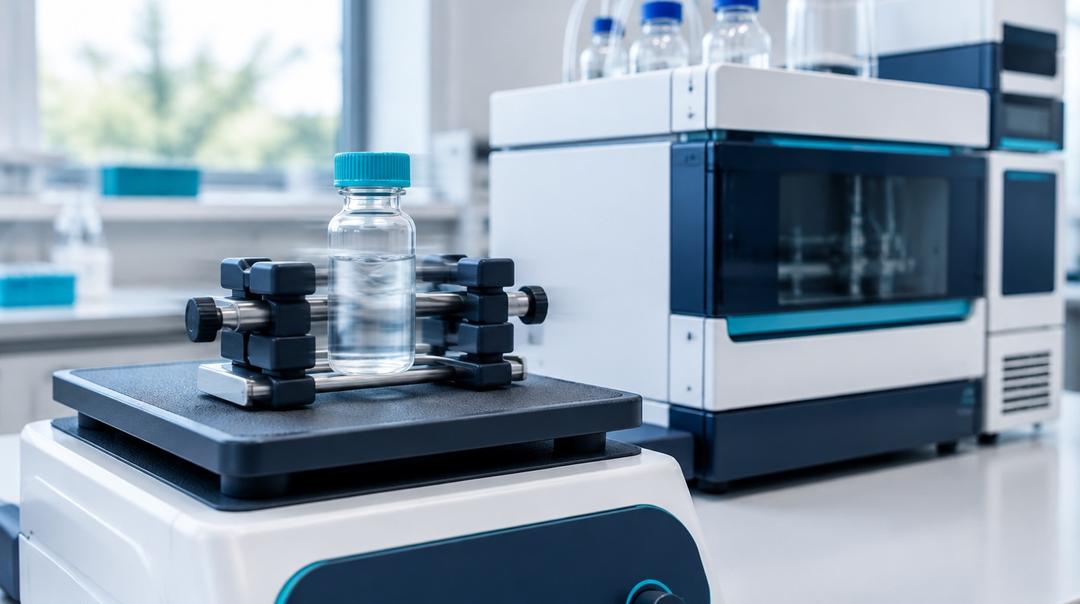
The practical advice not to shake a vial is already in handling research peptides in the laboratory. This page is about why that advice exists, how agitation stress is studied deliberately in formulation work, and why a single clean result does not settle the question for any peptide but the one tested.
Is it the shear force that damages peptides?
Mostly not. The intuitive picture — molecules torn apart by the violence of shaking — does not survive measurement. In one study, two concentrated monoclonal antibody formulations were exposed to shear rates up to 250,000 per second, far beyond anything a vial experiences, and shear alone did not cause aggregation. The authors calculated that the forces shear applies to a protein are tiny compared with the force expected at an air–water interface, and concluded that bubble entrainment, adsorption to solid surfaces and particulate contamination are more likely culprits (Bee and colleagues, PMID 19370772).
That work used large proteins, but the physics of the interface applies to anything surface-active. Air is hydrophobic. A molecule with hydrophobic side chains lowers its energy by placing them towards the air, and in doing so may unfold, reorient or pack against its neighbours. Shaking matters because it continually tears the interface open, folds adsorbed material back into the bulk solution, and replaces it with fresh surface. Foam is the extreme case: a large interfacial area loaded with adsorbed peptide.
What actually happens to a peptide at an interface?
Four outcomes are possible, and they can occur together.
Aggregation. Molecules concentrated and partly unfolded at the interface associate with each other. Aggregates range from reversible small oligomers to insoluble particles, and the review literature treats interfaces and agitation as standard induction factors for aggregation alongside temperature and freeze–thaw (Mahler and colleagues, PMID 18823031; Wang, PMID 15652195).
Fibril formation. Some peptides, once nucleated, grow into ordered amyloid-like fibrils. Insulin is the classic example: a study of its aggregation on agitation in the presence of hydrophobic surfaces examined how agitation rate, interfacial interactions and concentration governed the rate of aggregation, and found it proceeded in two stages (Sluzky and colleagues, PMID 1946348).
Adsorption to the container. Material can leave solution without aggregating at all, by sticking to glass or plastic. For three cationic membrane-active peptides, 90 per cent or more could be lost from solution to container walls at typical experimental concentrations (Kristensen and colleagues, PMID 25932639). Agitation increases contact between solution and wall.
Interaction with excipients. Surfactants are added to protein formulations precisely to occupy the interface first. In peptides the picture is less tidy: under agitation, liraglutide aggregated more extensively in the presence of polysorbate 80 and poloxamer 188, semaglutide changed little, and glucagon fibrillated regardless of surfactant (Hamada and colleagues, PMID 42679928). The same protective strategy helped one molecule and hurt another.
Covalent chemistry can also be affected — agitation introduces air, and with it oxygen — but for most peptides the physical routes above are the ones agitation studies are designed to detect.
Which peptides are most sensitive to shaking?
There is no universal ranking, but the features that raise susceptibility are well understood.
| Feature | Why it matters | Examples from the literature |
|---|---|---|
| Amphipathic structure | Strong tendency to adsorb at air–water and solid interfaces | GLP-1 analogues such as liraglutide |
| Intrinsic fibril-forming propensity | A nucleus formed at the interface can seed growth in the bulk | Glucagon, insulin |
| Self-association into oligomers | Interfacial stress can disrupt the native oligomer and redirect it to aggregates | Liraglutide, in contrast with semaglutide |
| Net positive charge with hydrophobic residues | Binding to negatively charged glass surfaces | Cationic membrane-active peptides |
| Low concentration | A larger fraction of total material sits at surfaces | Applies broadly |
The converse is a reasonable hypothesis rather than an established fact: very short, hydrophilic sequences with little structure to lose — a tripeptide, for instance — have less to unfold and weaker surface activity, and may tolerate agitation better. But "may" is the honest word. Robustness has to be shown for the molecule in question, in its formulation, with methods that could have detected damage. Cloudiness in a long, lipidated amylin analogue, and what it can and cannot mean, is discussed in cagrilintide cloudiness and aggregation.
Formulation details matter as much as sequence. In stress testing of an IgG1 antibody, the headspace in the vial strongly influenced how much damage shaking caused, temperature affected shaken but not stirred samples, and stirring and shaking produced qualitatively different aggregate species (Kiese and colleagues, PMID 18240293). The same molecule, shaken in a fuller vial, can give a different answer.
How is agitation damage detected?
The central difficulty is that aggregates span an enormous size range, from dimers a few nanometres across to particles visible to the eye, and no single method covers it. That is the explicit argument of a widely cited review asking whether any one measurement method is optimal for all aggregate sizes and types (Philo, PMID 17025274) — and of a commentary warning that subvisible particles, too large for chromatography and too small to see, were a gap that could compromise product quality (Carpenter and colleagues, PMID 18704929).
| Method | What it sees | What it misses |
|---|---|---|
| Visual inspection against light and dark backgrounds | Visible particles, haze, foam that persists | Everything below roughly the visible threshold |
| Turbidity or light scattering (static or dynamic) | Onset of soluble aggregates; size distribution in solution | Composition; it cannot say what the particles are |
| Subvisible particle counting (light obscuration, flow imaging) | Particle counts in the micrometre range | Small soluble oligomers |
| Size-exclusion chromatography (SEC) | Soluble dimers and oligomers separated from monomer | Insoluble aggregates filtered out before analysis; aggregates that dissociate on the column |
| Reversed-phase HPLC | Covalent changes and impurities resolved from the main peak | Most non-covalent aggregates, which dissolve in the organic, acidic mobile phase |
| LC-MS | Mass changes such as oxidation or cleavage | Changes that do not alter mass, including aggregation and conformation |
| Fluorescent dyes such as thioflavin T | Amyloid-like fibril structure | Amorphous aggregates |
| Quantitative recovery against a reference standard | Total material lost to adsorption or precipitation | The form the lost material took |
In the antibody study above, the authors concluded that an array of analytical methods was essential to cover the range of aggregate forms agitation produced (Kiese and colleagues, PMID 18240293). That is the general lesson.
Why one clean chromatogram doesn't settle it
The most common piece of evidence offered on this question is a before-and-after HPLC trace showing the same purity. It is a weaker test than it looks, for reasons specific to how the method works.
Reversed-phase conditions dissolve most aggregates. The sample is analysed in an acidified water–organic mobile phase that disrupts the non-covalent contacts holding a physical aggregate together. Material that was aggregated in the vial can elute as monomer.
Insoluble material never reaches the column. Samples are typically filtered or centrifuged before analysis to protect the column. A precipitate removed at that step simply does not appear.
Area-percent purity is a ratio. It reports the main peak as a share of everything detected. If a fraction of the peptide is lost to the vial wall or to a filtered-out precipitate, the remaining material can still be pure, and the purity figure unchanged, while the amount in solution has fallen. Only quantification against a reference standard reveals that loss.
One trace has no comparator. Without an unagitated control from the same lot, analysed in the same sequence, and without replicates, a small difference cannot be distinguished from method variability — and neither can the absence of one.
How each chromatographic method handles a peptide sample in general is set out in HPLC and LC-MS in peptide testing, and what a supplier's certificate can and cannot establish in what a certificate of analysis establishes.
What does a proper agitation study look like?
In formulation development, agitation is a deliberate stress test, run to find out whether a molecule is sensitive and which method detects the damage. A defensible design includes unagitated controls from the same lot, a defined shaker type, speed, duration and temperature, a stated fill volume and headspace, replicate vials, at least one method from each size range in the table above, and quantitative recovery against a reference standard. The result applies to that molecule, in that formulation and container, under that stress. It is evidence about a system, not a general rule about peptides.
If unexpected haze or particles appear in a laboratory solution, the observation itself needs investigating before any cause is assumed; a structured approach is in research peptide reconstitution problems. Nothing here is medical, dosing, or therapeutic guidance.
Frequently asked questions
Is shaking worse than vortexing or stirring? It depends on the molecule and the setup, and the answer is not intuitive. In one antibody study, stirring was more stressful than shaking, and the two produced different kinds of aggregates. What generally matters most is how much fresh air–liquid interface and foam the motion creates, and whether solution contacts hydrophobic surfaces such as a stir bar.
If the solution stays clear after shaking, is the peptide undamaged? Not necessarily. Soluble oligomers and subvisible particles are far smaller than the eye can resolve, and adsorption losses leave no visible trace at all. A clear solution rules out gross precipitation and nothing more. Confirming the absence of damage needs light scattering, particle counting or size-exclusion chromatography, and recovery against a reference standard.
Does foam mean the peptide has been damaged? Foam is peptide concentrated at a very large air–liquid interface, so it marks exactly the condition that drives interfacial aggregation in sensitive molecules. It is not proof of damage in itself — some molecules tolerate it — but material held in foam is unaccounted for, and its appearance is a reason to measure rather than assume.
Are small peptides immune to agitation damage? No peptide is immune by rule. Short, hydrophilic sequences with little structure plausibly tolerate agitation better than long amphipathic ones, but that is a hypothesis to test, not a finding to borrow. Even a robust peptide can be lost to container adsorption, which agitation increases and which purity testing alone does not detect.
Can a surfactant prevent agitation damage? Sometimes, and sometimes it makes things worse. Surfactants protect many proteins by occupying the interface first, but in a recent agitation study one peptide aggregated more with surfactant present, another changed little, and a third fibrillated regardless. Whether a surfactant helps has to be measured for each molecule and formulation.
Selected Research References
- Bee JS, Stevenson JL, Mehta B, et al. Response of a concentrated monoclonal antibody formulation to high shear. Biotechnology and Bioengineering. 2009;103(5):936-943. pubmed.ncbi.nlm.nih.gov/19370772
- Kiese S, Papppenberger A, Friess W, Mahler HC. Shaken, not stirred: mechanical stress testing of an IgG1 antibody. Journal of Pharmaceutical Sciences. 2008;97(10):4347-4366. pubmed.ncbi.nlm.nih.gov/18240293
- Mahler HC, Friess W, Grauschopf U, Kiese S. Protein aggregation: pathways, induction factors and analysis. Journal of Pharmaceutical Sciences. 2009;98(9):2909-2934. pubmed.ncbi.nlm.nih.gov/18823031
- Wang W. Protein aggregation and its inhibition in biopharmaceutics. International Journal of Pharmaceutics. 2005;289(1-2):1-30. pubmed.ncbi.nlm.nih.gov/15652195
- Sluzky V, Tamada JA, Klibanov AM, Langer R. Kinetics of insulin aggregation in aqueous solutions upon agitation in the presence of hydrophobic surfaces. Proceedings of the National Academy of Sciences of the USA. 1991;88(21):9377-9381. pubmed.ncbi.nlm.nih.gov/1946348
- Kristensen K, Henriksen JR, Andresen TL. Adsorption of cationic peptides to solid surfaces of glass and plastic. PLoS One. 2015;10(5):e0122419. pubmed.ncbi.nlm.nih.gov/25932639
- Hamada N, Wuelfing WP, Su Y, Ling J. Impact of surfactants on the stability of therapeutic peptides under interfacial stress. Journal of Pharmaceutical Sciences. 2026;115(11):104493. pubmed.ncbi.nlm.nih.gov/42679928
- Philo JS. Is any measurement method optimal for all aggregate sizes and types? The AAPS Journal. 2006;8(3):E564-E571. pubmed.ncbi.nlm.nih.gov/17025274
- Carpenter JF, Randolph TW, Jiskoot W, et al. Overlooking subvisible particles in therapeutic protein products: gaps that may compromise product quality. Journal of Pharmaceutical Sciences. 2009;98(4):1201-1205. pubmed.ncbi.nlm.nih.gov/18704929
All products sold by NovaEvum are for research and laboratory use only. They are not intended for human or animal consumption, diagnosis, treatment, cure, or prevention of any disease.