
Testing, quality and handling
Disulfide Bonds in Peptides: Formation, Scrambling and How Connectivity Is Verified
NB
· 9 min read
A summary of published research, with citations. It states no amount, schedule or route, and makes no claim about what this compound does in a person. Supplied for laboratory research use only.
On this page · 6 sections
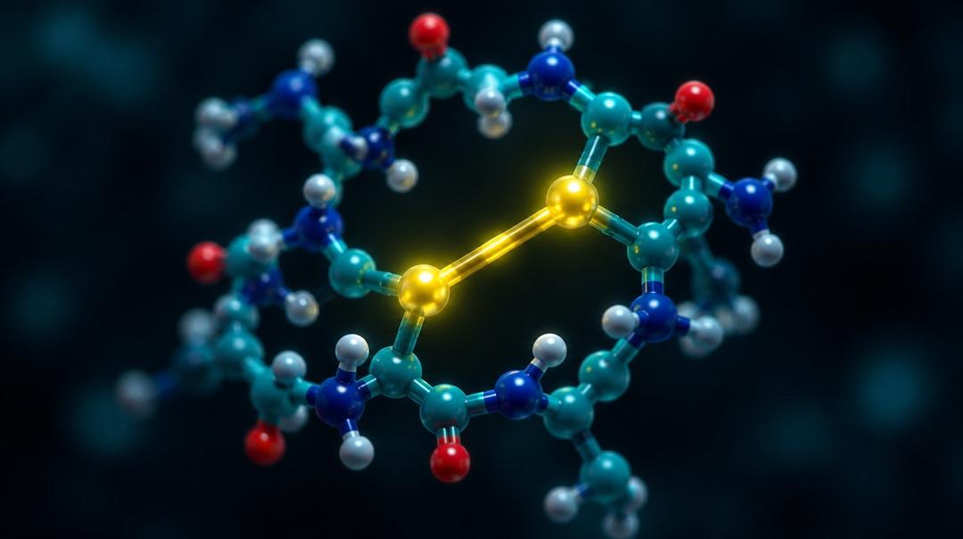
A disulfide bond is a covalent link between the sulfur atoms of two cysteine residues, formed by oxidation and broken by reduction. Within one molecule it staples the chain into a loop; between two molecules it joins them into a dimer. A peptide with four cysteines has three possible pairings, all of identical mass, so an intact mass measurement cannot say which one is present. Establishing connectivity requires cleaving the chain between the cysteines and weighing the linked fragments.
How does a disulfide bond form?
By oxidation of two thiol groups, with the loss of two hydrogen atoms. Dissolved oxygen will do it slowly; it goes faster at alkaline pH, where the thiol is deprotonated to the more reactive thiolate, and faster again in the presence of trace metals. The reverse reaction needs a reducing agent.
Two consequences follow immediately. A free cysteine in a peptide is not a stable feature of the molecule — it is a reactive group waiting for an oxidant, and that reaction is among the degradation routes catalogued in peptide degradation pathways. And the mass change is small: about 2 Da lighter than the reduced form, which is within the range of ordinary isotopic confusion unless the measurement is good.
Intramolecular and intermolecular bonds compete, and concentration decides between them. At low concentration a cysteine is most likely to meet another cysteine on its own chain; at high concentration it meets one on a neighbouring molecule, giving a covalent dimer. This is why oxidative folding of cysteine-rich peptides is conventionally run dilute. Moroder and colleagues review the alternatives: let the peptide fold oxidatively and hope it finds the native pairing, or build each bridge deliberately using orthogonal cysteine protecting groups removed one pair at a time (PMID 8785364).
Góngora-Benítez, Tulla-Puche and Albericio's review covers the whole territory for therapeutic peptides, from the synthetic strategies to what the bonds contribute once they are in place (PMID 24446748).
What does a disulfide bond do to a peptide?
It constrains the shape. A bridge removes most of the conformational freedom between the residues it links, which is the same mechanism that makes any cyclic peptide more rigid — see cyclic peptides explained. For small peptides the bridge is frequently the only thing giving the molecule a defined structure at all.
It also raises resistance to proteolysis, partly by rigidity and partly because a cut inside a disulfide loop does not separate the fragments: they stay tethered. Many of the most protease-resistant natural peptides — venom toxins, plant defence peptides, antimicrobials — are dense with cysteines for this reason.
Three well-known molecules illustrate the range:
| Peptide | Disulfide arrangement | Status |
|---|---|---|
| Oxytocin | One intramolecular bridge closing a ring, with a short tail outside it | A long-established licensed medicine |
| Octreotide | One intramolecular bridge closing an octapeptide ring; also carries two D-residues (PMID 6128648) | Licensed somatostatin analogue, reviewed in the clinical literature (PMID 8532003) |
| Insulin | Two chains joined by two interchain bridges, plus one intrachain bridge | Licensed medicine; the classical case where connectivity is the molecule |
Insulin makes the point most clearly. Its two chains are separate molecules held together by disulfides. Get the pairing wrong and you do not have a variant of insulin, you have a different substance.
Whether a disulfide survives depends on where the peptide is. The cytosol is a reducing environment, so intracellular disulfides are rare and mostly transient, while extracellular proteins keep theirs. The same logic applies to the glutathione couple, where the reduced and oxidised forms differ by exactly this chemistry — see reduced versus oxidised glutathione.
What is disulfide scrambling?
Scrambling is the rearrangement of correct pairings into incorrect ones, without any change to the molecule's atoms. A thiolate attacks an existing disulfide, displaces one of its partners, and the displaced thiolate attacks another bond in turn. One free thiol is enough to start the chain, and alkaline pH accelerates it by increasing the thiolate population.
The products are isomers. They have the same sequence, the same formula and the same mass as the native molecule, and they differ only in which sulfur is bonded to which. Chang's group prepared the full set of scrambled isomers of denatured epidermal growth factor deliberately, as a way of studying folding (PMID 12018622), and the same work established how readily these species form under denaturing conditions.
For a peptide with multiple cysteines, the number of possible pairings grows quickly: four cysteines give three arrangements, six give fifteen. A synthesis that folds oxidatively without directing the pairing can produce several of them, and a chromatogram may or may not separate them. This is the clearest case of the limit stated in HPLC and LC-MS testing: identical mass, possibly identical retention, and a different molecule.
Reduction is the other direction, and it is easy to cause accidentally. Common laboratory reductants open disulfides readily, as do the thiol-containing components of many biological buffers. A peptide analysed after contact with a reducing agent is a different species from the one that went in.
How is disulfide connectivity verified?
The general strategy is to cut the chain between the cysteines and see which pieces remain attached to which.
Non-reduced versus reduced comparison. Gorman, Wallis and Pitt's review sets out the standard approach: digest the protein with its disulfides intact, then compare the masses of the digest before and after reduction. A peak that disappears on reduction and is replaced by two smaller ones was a disulfide-linked pair, and the two masses identify the fragments (PMID 12476442).
The right protease. The same review makes the case for pepsin, whose acidic pH optimum is where disulfide rearrangement is least likely, and whose broad specificity gives cleavage between most cysteines. Working at low pH is not a detail — it is what stops the analysis from scrambling the sample it is measuring. Its drawback is ragged, overlapping products that complicate the spectra.
Isotope labelling of the cut ends. Digesting in a solvent containing heavy-oxygen water marks each newly formed carboxyl terminus, giving disulfide-linked products a distinctive isotope signature that identifies them among everything else in the digest (PMID 12476442).
Tandem mass spectrometry. Where a fragment carries more than one disulfide, fragmenting it further resolves the arrangement. Durand and colleagues developed a derivatisation route — adding methoxy to the disulfide before tandem analysis — specifically to assign linkage patterns among regio-isomers that are otherwise indistinguishable (PMID 28194735). The general fragmentation principles are in peptide sequencing by tandem mass spectrometry; disulfides complicate them, because a bridge holds fragments together that the instrument has already cleaved.
Free thiol quantification. Ellman's reagent, described in 1959, reacts with free sulfhydryl groups to give a coloured product measurable by absorbance (PMID 13650640). It does not tell you where the bonds are, but it tells you how many cysteines are not in one — which is often the first question, since a peptide that should be fully oxidised and shows free thiol has either failed to fold or has been partly reduced.
Composition after oxidative hydrolysis. Cysteine is one of the residues ordinary acid hydrolysis destroys, so compositional work converts it first to a stable derivative, classically by oxidation to cysteic acid. That route is part of amino acid analysis, and it determines total cysteine rather than how it was paired.
What should a certificate say about disulfides?
For a cysteine-containing peptide, three things that most research-chemical certificates omit.
First, whether the material is the oxidised or reduced form, since the two are different substances with a small mass difference and quite different behaviour. Second, free thiol content, which is the cheap check that the oxidation went to completion. Third, for anything with more than two cysteines, evidence of the connectivity — which means a digest comparison, not an intact mass.
A report showing only chromatographic purity and an intact mass for a multi-cysteine peptide has established that a species of the right weight is the main component. It has not excluded the scrambled isomers, because they weigh the same. That is a specific, nameable gap rather than a general complaint, and it belongs with the other limits in what a certificate of analysis establishes and the broader survey in third-party peptide testing in Canada. Nothing here is medical, dosing, or therapeutic guidance.
Frequently asked questions
How much does a disulfide bond change a peptide's mass? Forming one removes two hydrogen atoms, so the oxidised form is about 2 Da lighter than the reduced form. That is a small difference, and distinguishing it reliably needs a measurement good enough to resolve the isotope pattern. It is also the entire mass evidence available, since every possible pairing of the same cysteines gives the same number.
Can HPLC separate scrambled disulfide isomers? Sometimes, because different pairings fold the molecule differently and can expose different surfaces. There is no guarantee, and two isomers can co-elute as a single peak. Separation is a property of the particular molecule and method, not something a chromatogram can be assumed to deliver.
What causes a peptide to form dimers through disulfides? A free cysteine thiol on one molecule meeting one on another, under oxidising conditions. Concentration is the main lever: intramolecular pairing is favoured when molecules are far apart, intermolecular pairing when they are close. Alkaline pH and trace metal ions accelerate both.
Is a reduced peptide the same compound as the oxidised one? No. They differ by two hydrogen atoms and, more importantly, by structure: the oxidised form is constrained into a ring and the reduced form is a flexible chain. Where a molecule's activity depends on the bridge, the reduced form is a different substance with different properties.
Why use pepsin for disulfide mapping rather than trypsin? Because pepsin works at acidic pH, where thiolate concentration is low and disulfide exchange is slowest, so the digestion is less likely to rearrange the bonds it is meant to report. Its broad specificity also increases the chance of cleaving between every pair of cysteines. The cost is a complex digest of overlapping products.
Selected Research References
- Góngora-Benítez M, Tulla-Puche J, Albericio F. Multifaceted roles of disulfide bonds: peptides as therapeutics. Chemical Reviews. 2014;114(2):901-926. pubmed.ncbi.nlm.nih.gov/24446748
- Gorman JJ, Wallis TP, Pitt JJ. Protein disulfide bond determination by mass spectrometry. Mass Spectrometry Reviews. 2002;21(3):183-216. pubmed.ncbi.nlm.nih.gov/12476442
- Moroder L, Besse D, Musiol HJ, et al. Oxidative folding of cystine-rich peptides vs regioselective cysteine pairing strategies. Biopolymers. 1996;40(2):207-234. pubmed.ncbi.nlm.nih.gov/8785364
- Chang JY, Li L. The disulfide structure of denatured epidermal growth factor: preparation of scrambled disulfide isomers. Journal of Protein Chemistry. 2002;21(3):203-213. pubmed.ncbi.nlm.nih.gov/12018622
- Durand KL, Tan L, Stinson CA, et al. Assigning peptide disulfide linkage pattern among regio-isomers via methoxy addition to disulfide and tandem mass spectrometry. Journal of the American Society for Mass Spectrometry. 2017;28(6):1099-1108. pubmed.ncbi.nlm.nih.gov/28194735
- Ellman GL. Tissue sulfhydryl groups. Archives of Biochemistry and Biophysics. 1959;82(1):70-77. pubmed.ncbi.nlm.nih.gov/13650640
- Bauer W, Briner U, Doepfner W, et al. SMS 201-995: a very potent and selective octapeptide analogue of somatostatin with prolonged action. Life Sciences. 1982;31(11):1133-1140. pubmed.ncbi.nlm.nih.gov/6128648
- Lamberts SW, van der Lely AJ, de Herder WW, et al. Octreotide. New England Journal of Medicine. 1996;334(4):246-254. pubmed.ncbi.nlm.nih.gov/8532003
All products sold by NovaEvum are for research and laboratory use only. They are not intended for human or animal consumption, diagnosis, treatment, cure, or prevention of any disease.